
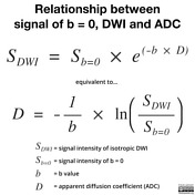
Web This is known as the b0 image. DWI is done to determine the rate of molecular diffusion in different areas of the body.

Apparent Diffusion Coefficient Radiology Reference Article Radiopaedia Org
Certain illnesses show restrictions of diffusion for example demyelinization and cytotoxic edema.
. In DWI the optimal b value is 600 smm 2. Web ADC values are typically calculated from a set of MR images obtained with varying degrees of diffusion weighting b-values using nonlinear regression. Widest Range Of Refurbished MRI Equipment We Sell Top MRI Machine Brands.
We tested the correlations between cortical microarchitecture and diffusion metrics computed from standard 1000 smm2 high 3000 smm2 to very high 5000 smm2 b-value dMRI. Web Effects of magnetic field strength and b value on the sensitivity and specificity of quantitative breast diffusion-weighted MRI. The aim of our study was to determine how many b-values are necessary to reliably calculate ADC maps.
Strength duration and the period between diffusion gradients. Next the ease with which water can diffuse is assessed in various directions. Mean ADC value is 13 higher in total by additional use of b0 and b50 smm 2 in multiple b-value combinations.
Web The b-value reflects the intensity and sensitivity of diffusion restriction and is like the torque of a cars engine. Web The degree of diffusion weight in g correlates with the strength of the diffusion gradients characterized b y the b - value which is a function of the gradient related parameters. Web The b value is used in MRI in the context of Diffusion Weighted Imaging DWI.
Certa in illnesses show restrictions of diffusion for example demyel in ization and cytotoxic edema. A total of 124 consecutive men with suspected prostate cancer PCa underwent diagnosis prostate MRI on a 30 T MR. To compare computed high b-value diffusion-weighted images c-DWI derived from low b-value DWI images and acquired high b-value DWI a-DWI in overall image quality and prostate cancer detection rate.
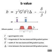
Web b value measures the degree of diffusion weighting applied thereby indicating the amplitude G time of applied gradients δ and duration between the paired gradients Δ and is calculated as. Web The degree of diffusion weighting correlates with the strength of the diffusion gradients characterized by the b-value which is a function of the gradient related parameters. Multiple b values of 600 smm 2 and higher are recommended to differentiate between benign and malignant abdominal lesions.
Strength duration and the period b etween diffusion gradients. However there is no agreement concerning the number of images needed for ADC calculation. The lesion ADCnormal parenchyma ADC ratio is.
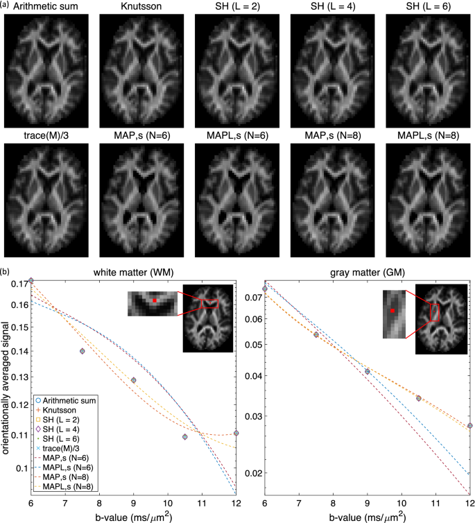
B γ² G² δ² Δδ3. A typical series of b-values would be 0 50 150 800 and 1600. Web Purpose In diffusion MRI dMRI it remains unclear to know how much increase of b-value is conveying additional biological meaning.
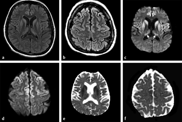
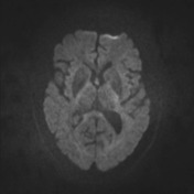
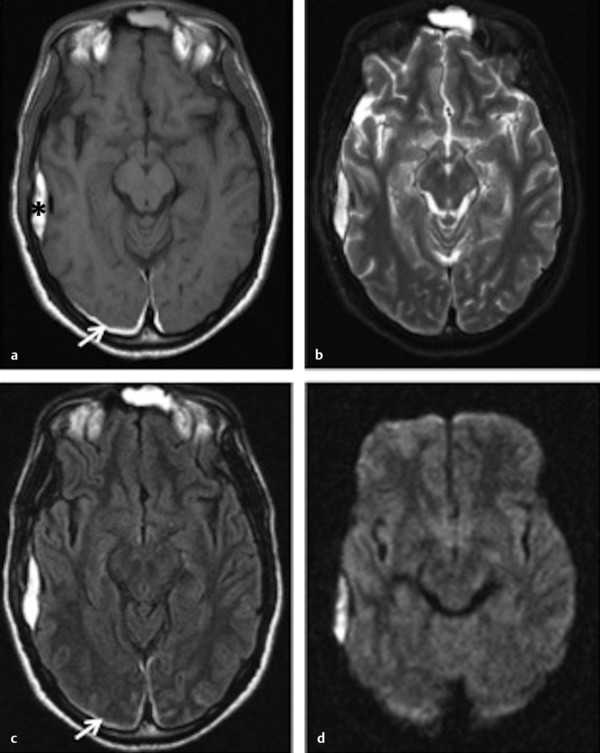
In general in healthy tissue molecules of water and other chemicals are not stationary but moving about. YOU MUST have a b-value of at least 1400. Web High b-value diffusion-weighted MRI of normal brain Brain DW images obtained at b 3000 appear significantly different from those obtained at b 1000 reflecting expected loss of signal from all areas of brain in proportion to their ADC values.
The minimum is 3 orthogonal directions X Y and Z and we will use this for the rest of this explanation. Ad We Sell Used Refurbished MRI Equipment Check Out Our Buyers Guide Here. Authors Mohammad Eghtedari 1 Jingfei Ma 2 Patricia Fox 3 Inanc Guvenc 4 Wei T Yang 5 Basak E Dogan 6 Affiliations 1 Department of Radiology University of California San Diego San Diego CA USA.
Web High b-value diffusion MR imaging provides an enhanced contrast toward different cellular components 1 2 when compared with clinical diffusion MRI b 1000 smm 2Combined with an appropriate model the analysis of low and high b-value diffusion images may provide a comprehensive tissue characterization enabling improved. This is done by applying a strong gradient symmetrically on either side of the 180-degree pulse. In DWI we recommend the use of b-values of 0 and 800 smm 2 as two b-values or b0 50 600 800 and 1000 smm 2 as multiple b-values for distinguishing between benign and malignant liver lesions.
The higher the b-value the more sensitive the sequence is to diffusion restriction.
Diffusion Weighted Imaging Radiology Reference Article Radiopaedia Org
2
Diffusion Weighted Imaging In Hemorrhage Radiology Key

Principles Of Diffusion Tensor Imaging And Its Applications To Basic Neuroscience Research Neuron
2
Computing The Orientational Average Of Diffusion Weighted Mri Signals A Comparison Of Different Techniques Scientific Reports

Diffusion Weighted Imaging Radiology Reference Article Radiopaedia Org
Diffusion Weighted Imaging Radiology Reference Article Radiopaedia Org

Diffusion Weighted Imaging Radiology Reference Article Radiopaedia Org
Tensor Valued Diffusion Encoding For Diffusional Variance Decomposition Divide Technical Feasibility In Clinical Mri Systems Plos One
Tensor Valued Diffusion Encoding For Diffusional Variance Decomposition Divide Technical Feasibility In Clinical Mri Systems Plos One

Diffusion Weighted Imaging Radiology Reference Article Radiopaedia Org
Diffusion Weighted Imaging Radiology Reference Article Radiopaedia Org

Diffusion Weighted Imaging In Acute Ischemic Stroke Radiology Reference Article Radiopaedia Org
Diffusion Weighted Imaging In Hemorrhage Radiology Key

Hyperpolarised 13c Mri Identifies The Emergence Of A Glycolytic Cell Population Within Intermediate Risk Human Prostate Cancer Nature Communications
2

Apparent Diffusion Coefficient Radiology Reference Article Radiopaedia Org
Diffusion Weighted Imaging In Hemorrhage Radiology Key